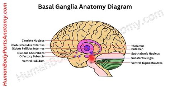
What are basal ganglia?
The basal ganglia are a set of subcortical nuclei in the cerebrum that are involved in the integration and selection of voluntary behaviour. The striatum, the major input station of the basal ganglia, has a key role in instrumental behaviour — learned behaviour that is modified by its consequences.
What does T1 hyperintense mean?
T1 signal hyperintensity may correspond to intracellular and extracellular methemoglobin. It may also be seen during the chronic stage of a clot or hemorrhage, when sedimentation of the blood cells produces a distinctive fluid-debris level within the lesion.
What disease is associated with basal ganglia?
Parkinson’s. Parkinson’s is the most notorious disease of the basal ganglia. Classic clinical symptoms include bradykinesia, resting tremor, postural instability, and shuffling gait. This disease is a result of neurodegeneration of the SNpc dopaminergic neurons.
Is basal ganglia involved in memory?
Extensive evidence now indicates a role for the basal ganglia, in particular the dorsal striatum, in learning and memory. One prominent hypothesis is that this brain region mediates a form of learning in which stimulus-response (S-R) associations or habits are incrementally acquired.
What is T2 and flair Hyperintensities?
Focal hyperintensities in the subcortical white matter demonstrated by T2-weighted or FLAIR images are a common incidental finding in patients undergoing brain MRI for indications other than stroke. They are indicative of chronic microvascular disease.
What does Flair hyperintensity mean?
There are a variety of MRI sequences or imaging patterns used (ie. T1, T2 or FLAIR) to highlight or suppress different types of tissue so that abnormalities can be detected. Hyperintensity on a T2 sequence MRI basically means that the brain tissue in that particular spot differs from the rest of the brain.
What is Flair hyperintensity on MRI?
White matter hyperintensities (WMHs) are clinically silent abnormalities visible in deep or periventricular white matter on CT or MRI. They are particularly apparent on FLAIR MRI, which is a T2-weighted sequence where the CSF signal is suppressed.
