What does tonsillar fossa mean?
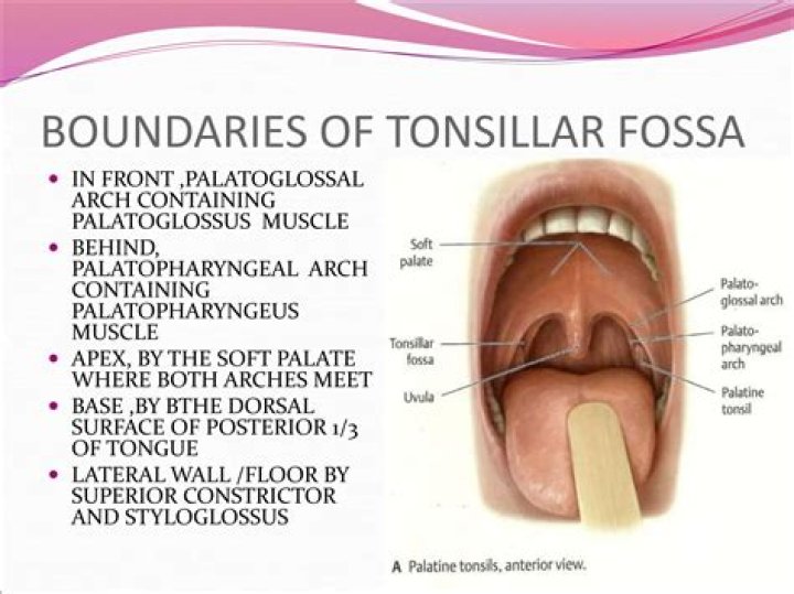
The tonsillar fossa (Tonsillar sinus; ; Tonsillar bed) is the depression between the palatoglossal and palatopharyngeal arches (as well as the triangular and semilunar folds) occupied by the palatine tonsil.
What is right tonsillar fossa?
The tonsillar fossa (or tonsillar sinus) is a space delineated by the triangular fold (plica triangularis) of the palatoglossal and palatopharyngeal arches within the lateral wall of the oral cavity. . In many cases, however, this sinus is obliterated by its walls becoming adherent to the palatine tonsils.
Where are tonsils palpated?
Palpate glossotonsillar sulcus (where palatine tonsil meets posterior base of tongue). Slide finger in gently from retromolar trigone or anterior pillar. Assess palatine tonsils for symmetry in size and color. Document tonsil size (Brodsky score).
What is the tonsillar region?
The tonsils (palatine tonsils) are a pair of soft tissue masses located at the rear of the throat (pharynx). Each tonsil is composed of tissue similar to lymph nodes, covered by pink mucosa (like on the adjacent mouth lining). The tonsils are part of the lymphatic system, which helps to fight infections.
What is the tonsillar pillar?
The anterior tonsillar pillar is formed by the palatoglossus muscle, and the posterior pillar is formed by the palatopharyngeus muscle. The anterior pillar is posterior to the retromolar trigone. The soft palate serves as the roof of the oropharynx and the floor of the nasopharynx.
What is tonsillar capsule?
Description. From the pharyngeal side, the palatine tonsils are covered with a stratified squamous epithelium (the tonsillar capsule), whereas a fibrous capsule links them to the wall of the pharynx. Through the capsule pass trabecules that contain small blood vessels, nerves and lymphatic vessels.
What is tonsillar pillar?
Posterior tonsil pillar This is the fold of tissue just behind the tonsils. It is created by the palatopharyngeus muscle which extends from the soft palate to the lateral wall of the pharynx.
How do you palpate tonsillar lymph nodes?
Tonsillar nodes: At the angle of Mandible. Deep cervical lymph nodes should be palpated, one side at a time. Gently bend the patient’s head forward and roll your fingers over the deeper muscles along the carotid arteries. To feel Scalene nodes roll your fingers gently behind the clavicles.
How do you palpate tonsillitis?
Gently feeling (palpating) your child’s neck to check for swollen glands (lymph nodes) Listening to his or her breathing with a stethoscope. Checking for enlargement of the spleen (for consideration of mononucleosis, which also inflames the tonsils)
What is the function of tonsil?
The tonsils are part of the body’s immune system. Because of their location at the throat and palate, they can stop germs entering the body through the mouth or the nose. The tonsils also contain a lot of white blood cells, which are responsible for killing germs.
What is behind the tonsil?
What are adenoids? Adenoids are a patch of tissue that is high up in the throat, just behind the nose. They, along with the tonsils, are part of the lymphatic system.
What is the area behind the uvula called?
The nasopharynx is the portion of the pharynx that is posterior to the nasal cavity and extends inferiorly to the uvula. The oropharynx is the portion of the pharynx that is posterior to the oral cavity.
