What is scanning probe microscopy used for?
Scanning probe microscopy (SPM) is a method of sample surface observation that uses a physical probe to interrogate a specimen rather than light. This provides a wealth of information that cannot be obtained via light microscopy.
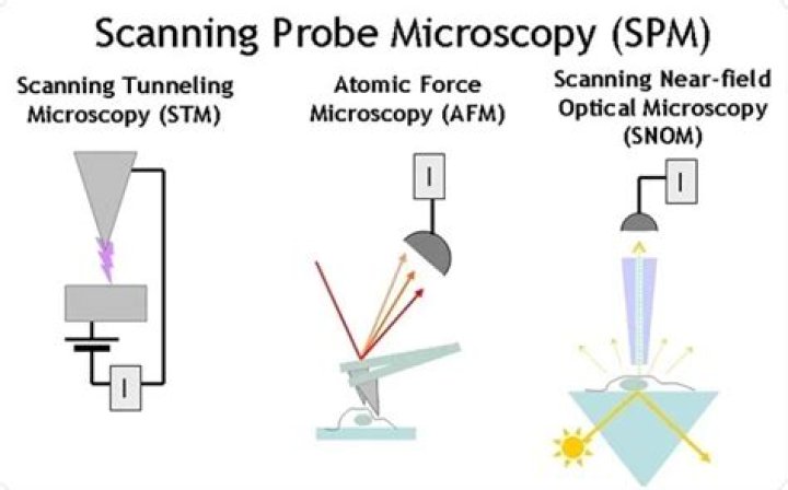
What are the types of scanning probe microscope?
There are various other types of scanning probe microscopes including:
- Ballistic electron emission microscopy (BEEM)
- Chemical force microscopy (CFM)
- Conductive atomic force microscopy (C-AFM)
- Electrochemical scanning tunneling microscopy (ECSTM)
- Electrostatic force microscopy (EFM)
- Fluidic force microscopy (FluidFM)
What is the main advantages of scanning probe microscopic techniques?
The main advantages of scanning probe microscopy are as follows: – High locality due to the probe-surface interaction; – The probe may be used to modify the structure of a sample’s surface; – The probe can be used in vacuum, in air and in liquid environments.
Which property is measured with a scanning probe microscope?
SPMs are designed to measure local properties, such as friction, height, and magnetism to acquire an image with a probe, the SPM raster scans the probe over a small area of the sample, measuring the local property simultaneously.
What can AFM measure?
AFM is used to measure and localize many different forces, including adhesion strength, magnetic forces and mechanical properties. AFM consists of a sharp tip that is approximately 10 to 20 nm in diameter, which is attached to a cantilever. The tapping mode is the recommended mode that is commonly used for AFM imaging.
What are the basic parts of any scanning probe microscopy SPM )?
1: The main components of a scanning probe microscope (SPM) used for surface analysis, which includes the piezo-tube actuator, the probe tip, and the sample.
What are the three types of AFM scanning?
AFM has three differing modes of operation. These are contact mode, tapping mode and non-contact mode.
What are the disadvantages of scanning probe microscope?
The disadvantages are that it cannot be used for solid–solid or liquid–liquid surfaces, as the maximum image size is smaller. The idea of topography, electrical, as well as magnetic properties of the nanomaterials is acquired using SPM. Using SPM information can also be transferred to sample.
How is a scanning probe microscope different from an electron microscope?
Electron microscopes use electromagnetic or electrostatic lenses and a beam of charged particles (instead of light) to view particles of size in the nanometer scale, e.g., atoms. Scanning probe microscopy was developed in the 1980s to study atomic surfaces at nanoscale resolution. These microscopes don’t use lenses.
How does a scanning electron microscope work?
The SEM is an instrument that produces a largely magnified image by using electrons instead of light to form an image. A beam of electrons is produced at the top of the microscope by an electron gun. Once the beam hits the sample, electrons and X-rays are ejected from the sample.
What is AFM analysis?
Atomic Force Microscopy (AFM) analysis provides images with near-atomic resolution for measuring surface topography. AFM is also referred to as Scanning probe microscopy. It is capable of quantifying surface roughness of samples down to the angstrom-scale.
