What is V1 and V2 in ECG?
V1, V2 = RV. V3, V4 = septum. V5, V6 = L side of the heart. Lead I = L side of the heart. Lead II = inferior territory.
Why is V1 and V2 negative in ECG?
In right chest leads V1 and V2, the QRS complexes are predominantly negative with small R waves and relatively deep S waves because the more muscular left ventricle produces depolarization current flowing away from these leads.
What is RSR pattern in V1?
When the QRS is ≥120 ms, usually this pattern corresponds to advanced right bundle branch block (RBBB) and/or right ventricular hypertrophy; or to some types of ventricular preexcitation (WPW pattern) [1], [2]. The ECG pattern of Rsr′ in leads V1-V2, with QRS < 120 ms, is a common electrocardiographic finding.
Where is V2 lead placed?
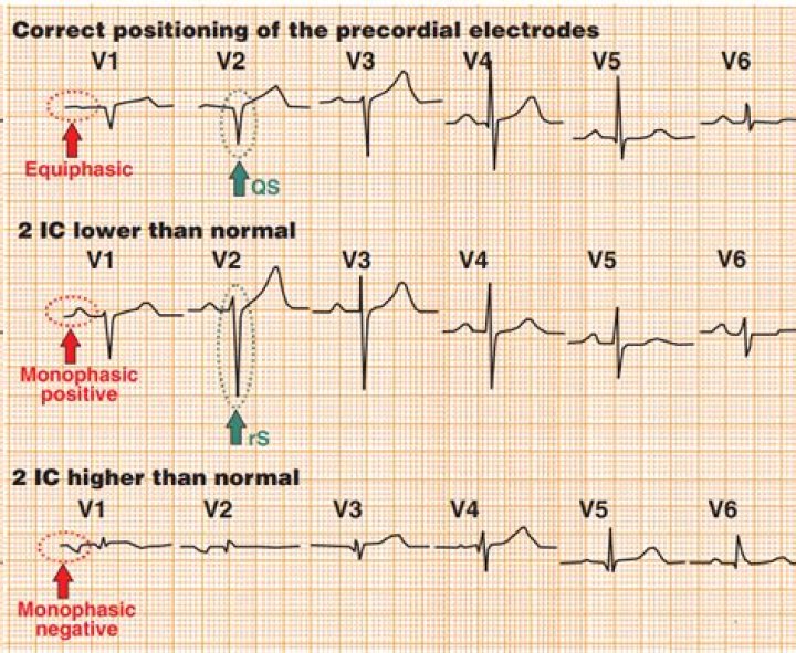
Precordial Lead Placement V1 is placed to the right of the sternal border, and V2 is placed at the left of the sternal border. Next, V4 should be placed before V3. V4 should be placed in the fifth intercostal space in the midclavicular line (as if drawing a line downwards from the centre of the patient’s clavicle).
Where do you put V1 and V2?
The proper location of V1 and V2 have not changed in many decades. They are located in the 4th intercostal space, just right and left, respectively, of the sternum. It is fairly easy to determine this spot using the angle of Louis as a landmark.
Is rSr in V1 normal?
The rSr’ pattern can be considered a normal variant due to delay in the activation of the basal part of the right ventricle (RV). It has been reported that an rSr’ pattern is a common finding in the general population.
What causes rSr in V1?
We often face this finding in asymptomatic and otherwise healthy individuals and the causes may vary from benign nonpathological variants to severe or life-threatening heart diseases, such as Brugada syndrome or arrhythmogenic right ventricular dysplasia.
Where do V1 V2 leads go?
V1 is placed to the right of the sternal border, and V2 is placed at the left of the sternal border.
