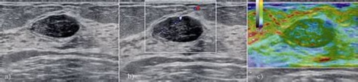
How does a fibroadenoma look on ultrasound?
Typically seen as a well-circumscribed, round to ovoid, or macrolobulated mass with generally uniform hypoechogenicity. Intralesional sonographically detectable calcification may be seen in ~10% of cases 2. Sometimes a thin echogenic rim (pseudocapsule) may be seen sonographically.
Can you see fibroadenoma on ultrasound?
While fibroadenomas may show up on a mammogram, they are often seen more clearly on an ultrasound of the breast. The diagnosis of a fibroadenoma can be confirmed by taking a sample of cells from the area for a pathologist to examine under the microscope.
Do fibroadenomas look different than cancer on ultrasound?
Cysts and carcinomas are better distinguished from fibroadenoma by ultrasound imaging; however, overlapping findings in nonhomogeneous fibroadenomas along with occasional calcification and noncircumscribed margins may mimic the findings in several other types of breast masses.
Are fibroadenomas round or oval?
Fibroadenomas are typically round, ovoid, or lobulated, with smooth margins; however, on early contrast-enhanced images, they may exhibit an irregular shape or margin resulting from the progression of enhancement.
What can fibroadenoma be mistaken for?
Fibroadenomas are almost always benign but there is a slight possibility of cancer, which is why a doctor must always perform a thorough examination. Sometimes the growths are misdiagnosed as an abscess or a fibrocystic condition, which calls for a different treatment process.
How do you diagnose a fibroadenoma?
The only way for a doctor to know for sure that it’s a fibroadenoma is through a biopsy, which means taking a sample of the lump to test in a lab. Based on the results of your examination and scan, your doctor will decide whether they need to get extra confirmation from a biopsy.
Where are fibroadenomas usually located?
A fibroadenoma is most often detected incidentally during a medical examination or during self examination, usually as a discrete solitary breast mass of 1 to 2 cm. Although they can be located anywhere in the breast, the majority are situated in the upper outer quadrant.
How can you tell the difference between a fibroadenoma and cancer?
Unlike a breast cancer, which grows larger over time and can spread to other organs, a fibroadenoma remains in the breast tissue. They’re pretty small, too. Most are only 1 or 2 centimeters in size. It’s very rare for them to get larger than 5 centimeters across.
Can a fibroadenoma look suspicious?
Fibroadenoma calcifications can range in morphology from round to coarse dystrophic to pleomorphic (Figure 2B-C). When beginning to calcify, fibroadenomas may appear suspicious, necessitating further imaging evaluation and biopsy.
When should a fibroadenoma be biopsied?
After 12 months, any persistent fibroadenoma should be excised. Management of a fibroadenoma (FA) in women older than 35 years of age. Our experience, and that reported by others,52 is that when offered the option of conservative management, most women will eventually prefer excisional biopsy.
Are fibroadenomas always biopsied?
RESULTS. Fibroadenomas of the breast are common, accounting for 50% of all breast biopsies performed. Physical examination, sonography, and fine needle aspiration are effective in distinguishing fibroadenomas from breast cancer.
